What Is an Isthmocele and How Does It Affect Pregnancy?
What Is an Isthmocele?
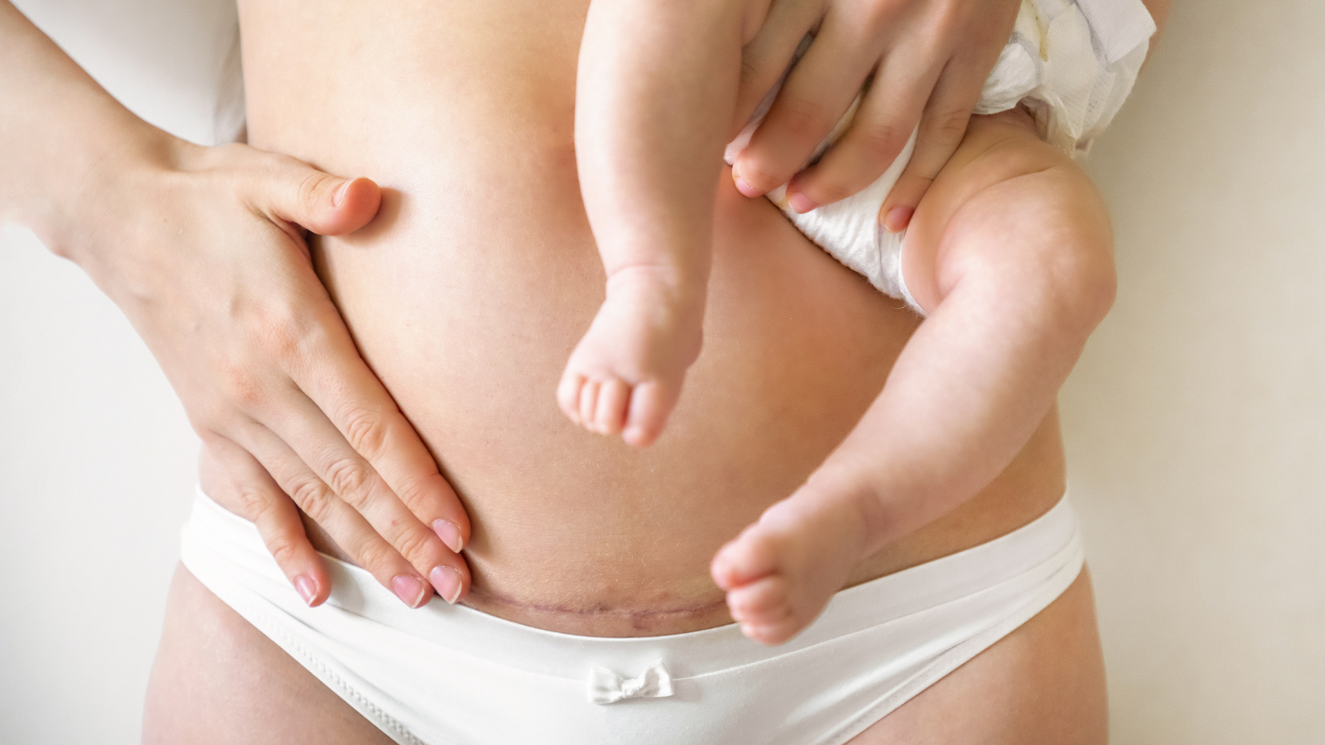
An isthmocele is a pouch-like defect that develops in the uterine wall following a caesarean delivery because the healing process is not fully completed. This pouching generally occurs in the anterior muscular layer of the uterus at the site of the caesarean incision.
An isthmocele may develop shortly after childbirth or many years later. Although it may not cause any symptoms in some women, it generally presents with complaints such as menstrual irregularities and vaginal discharge.
What Are the Symptoms of an Isthmocele?
The most common symptoms of an isthmocele include:
- Postmenstrual spotting: Brown, muddy-looking vaginal discharge
- Menstrual irregularities: Spotting between menstrual periods or delayed menstrual bleeding
- Groin pain: Pain experienced particularly during menstrual periods
- Pain during sexual intercourse: A sensation of significant discomfort or pain
- Vaginal discharge: An increase in vaginal discharge with a colour or consistency different from normal
- Painful menstruation: Severe cramps during menstrual periods
The accumulation of blood and fluid within the uterus after menstruation is one of the most notable symptoms of an isthmocele. This condition may impair sperm movement, making pregnancy more difficult and potentially causing infertility.
What Causes an Isthmocele?
Incomplete healing of the uterine wall following a caesarean delivery is the main cause of an isthmocele. The following factors may contribute to the development of this condition:
- Surgical techniques: The depth or angle of the incision made during the caesarean section
- Infections: The development of a uterine infection following childbirth
- Structural characteristics of the uterus: The uterine tissue of some women may be more susceptible to this condition
- Multiple caesarean deliveries: The risk increases in women who have undergone more than one caesarean delivery
Effects on Pregnancy
An isthmocele may lead to complications that directly affect pregnancy. In addition to impairing sperm movement and causing infertility, it may result in serious problems during pregnancy in some cases:
- Risk of miscarriage: The pouch-like defect in the uterus may make implantation of the embryo more difficult.
- Placenta previa: The placenta covers the cervix.
- Caesarean scar pregnancy: The embryo implants within the pouch-like defect, resulting in an ectopic pregnancy.
These complications may place the pregnancy at risk and require early intervention.
How Is an Isthmocele Diagnosed?
An isthmocele is generally diagnosed using the following imaging methods:
- Transvaginal ultrasonography: This is the first-line method used to visualise the pouch-like defect in the uterine wall.
- Hysteroscopy: An optical instrument used to examine the inside of the uterus in detail.
- Magnetic resonance imaging: This provides detailed imaging in more complex cases.
For the most accurate results, imaging is generally performed during the early proliferative phase of the menstrual cycle, immediately after menstruation has ended.
You may read this blog article for detailed information about hysteroscopy.
Treatment Methods for Isthmocele
The treatment plan is determined according to the patient’s symptoms and her desire to have children.
Medical Treatment
In patients with mild complaints, hormonal treatments may be used to regulate the menstrual cycle. However, this method generally provides only temporary relief.
Surgical Treatment
Surgical intervention provides a permanent solution for an isthmocele. Common surgical methods include:
Hysteroscopic Surgery
- The pouch-like area is corrected through the vaginal route using an instrument that visualises the inside of the uterus.
- The procedure generally takes 15–20 minutes, and the patient is discharged on the same day.
Laparoscopic Surgery
- A camera is inserted into the abdominal cavity through a small incision below the umbilicus.
- The pouch-like area is removed, and the healthy tissues are repaired with sutures.
- The procedure takes approximately one hour, and the patient can generally return to normal daily life one day later.
The preferred method depends on the size of the isthmocele and the patient’s pregnancy plans.
Vaginal Repair of an Isthmocele
- The pouch-like area is removed through the vaginal route, and the healthy tissues are repaired with sutures.
- The procedure takes approximately one hour, and the patient can generally return to normal daily life one day later.
Can an Isthmocele Be Prevented?
Ensuring appropriate healing following a caesarean delivery may reduce the risk of an isthmocele. However, it should be remembered that individual factors may also play a role. For this purpose:
- Postpartum follow-up appointments should not be neglected.
- Hygiene recommendations should be followed to prevent infections, and any infections should be treated.
- Appropriate surgical techniques should be used.
Frequently Asked Questions
Why Does an Isthmocele Make Pregnancy More Difficult?
Blood and fluid accumulating within the isthmocele pouch may prevent sperm from moving through the uterus. It may also make implantation of the embryo more difficult.
In Whom Is an Isthmocele More Common?
An isthmocele occurs in women who have undergone a caesarean delivery. It does not develop in women who have had a vaginal delivery because no incision is made in the uterus.
Can an Isthmocele Resolve Spontaneously?
No. An isthmocele generally requires surgical intervention. In mild cases, hormonal treatment may reduce the symptoms.
Can an Isthmocele Recur After Surgery?
With an appropriate surgical method and regular follow-up, the risk of recurrence is low.
Which Method Is Most Commonly Used to Diagnose an Isthmocele?
Transvaginal ultrasonography is the most commonly used and easily performed diagnostic method.
In conclusion, an isthmocele is a common condition, particularly among women who have undergone a caesarean delivery. With accurate diagnosis and appropriate treatment methods, the symptoms caused by this condition can be controlled and quality of life can be improved.
Receiving support from an experienced obstetrician and gynaecologist is highly important throughout this process. Prof. Dr Şadıman Kıykaç Altınbaş, a Specialist in Obstetrics and Gynaecology practising in Ankara, guides her patients as a physician specialising in the management of such conditions. You may contact us for further information.